Saturday, September 30, 2023

Are there potential problems in the latest study on antipsychotic medication reduction and discontinuation?


A study on antipsychotic medication reduction and discontinuation came out yesterday with fanfare.  The fanfare was basically because the principal investigator is a self-proclaimed critical psychiatrist with many criticisms of psychiatric medication and the results of her trial contradicted the primary hypothesis of the study and that was:

“Our hypothesis was that antipsychotic reduction would improve social functioning with only a small increase in relapse rate.”

Relapse rate in this case was defined as rehospitalization and the authors subsequently state that they thought a 10% rate of relapse would be “acceptable.”  The irony of this situation (ideology versus real world treatment) was not lost on anyone. Several people seemed to congratulate the authors on publishing results inconsistent with their ideology although the study was so embedded in the UK research infrastructure – I doubt that not publishing it would have been an option.

As a clinical trialist myself – the research seems to present several problems and creates several questions that could suggest that it was designed to optimize the likelihood that antipsychotic medication could be reduced and possibly discontinued. Before I get into those scenarios let me briefly summarize the results.  The paper is open access and can be downloaded as well as another paper that describes the research protocol.

In the study there were two arms an antipsychotic maintenance arm (N=127) and a reduction arm (N= 126). Diagnoses were taken from clinical information and the clinical staff had treatment responsibility for the patients.  In those patients who were randomized to dose reduction, a tapering protocol was suggested to the clinical staff and if it went well at some point the option for a more rapid taper or discontinuation was offered.  The research staff monitored the protocol. Baseline and outcome measure included a number of checklists to assess side effects, sexual side effects, positive and negative symptoms, quality of life, and social outcomes at the reassessment points.  Raters were blinded but the measures are essentially self report.  The ultimate result was that the risk of adverse outcomes was worse in the reduction arm with no associated improvement in social functioning.   

He are some potential issues that I noticed based on my experience in clinical trial design and on research review boards.

1.  Recruitment – described in the following:

“Participants were recruited from 19 National Health Service Trust mental health organisations across England. Potential participants were identified initially by clinical staff or recruited through advertisements placed in clinical settings and social media; those patients who expressed an interest in participating were sent further information”.

Not enough information. What did the advertisements say? Were subjects aware of who was running this trial and what the goal of the research was? Were the patients asked why they were interested in participating in this trial?  Were they asked what they think about taking a medication? Did the subjects have any exposure to the considerable press that the critical psychiatry group and the principal investigator generate?  Descriptions in the lay press have been demonstrated to have significant effects on perceived side effects – even to the point of creating a nocebo effect (6) – is there any reason to think that a group emphasizing side effects and minimizing any therapeutic effects might have a similar impact? If that is the case – how would it affect this trial?  

2.  Inclusion/Exclusion criteria

“Exclusion criteria included being considered by a clinician to pose a serious risk of harm to self or others were the individual to reduce their antipsychotic medication, being mandated to take antipsychotic medication under a section of the Mental Health Act, having been admitted to hospital or treated by a crisis service for a mental disorder within the last month, lacking capacity to consent, having insufficient spoken English, pregnancy, breastfeeding, and being involved in another trial of an investigational medical product; eligibility was assessed by researchers and confirmed by the Principal Investigator for the site.”

Practically all the exclusion criteria result in a population that may be more likely to discontinue antipsychotic medications with less difficulty. Consistent with this is the antipsychotic doses of both the reduction and maintenance arm of 300 mg chlorpromazine equivalents (on average).  According to the Maudsley Prescribing Guidelines (4) 300 mg chlorpromazine is considered the minimally effective dose of medication for relapsing schizophrenia. Whether this was a representative sample of the 4109 patients put forward for research by clinicians a comparison of the demographics and medication doses would have been of interest.  

Selection bias may also be evident in the Consort diagram (page 4).  After subjects consented to be contacted by the research team (N= 958) – a total of 562 declined participation. Was that because they did not want to take the chance of randomization to a medication reduction?

3.  Diagnoses – the diagnosis required was schizophrenia or non-affective psychoses with recurrent episodes. The diagnoses were taken from clinical records.  Considerable heterogeneity is introduced with the non-specific category of psychoses with an unpredictable course for which the concept of maintenance medication was not intended.  

4.  The Dose Reduction - 

The description of the dose reductions in the paper is confusing.  It starts out describing individualized reductions every 2 months based on starting doses but at some point states the patient is allowed to discontinue the medication if the dose reduction has been going well or reduce at a rate of the equivalent of 2 mg haloperidol/day.  2 mg/day of haloperidol is not a slow reduction and it is a departure from reduction every 2 months. Some of the authors here have written about antipsychotic withdrawal reactions – how is the more rapid dose reduction or optional abrupt discontinuation justified? 

4.  Safety Monitoring/Informed Consent: 

The more clinical trials I read (and I have read thousands) – the more I want to see the consent form that each patient signs. Some of the authors here continuously talk about medication side effects.  In fact – the principle investigator (PI) has stated that in her opinion that modern psychiatric medications work in a "drug centered" rather than a disease centered model by producing side effects like sedation, cognitive impairment, dysphoria, and loss of libido (5).  In that model, symptoms of mental illness are muted by side effects rather than effectively treated. The model essentially denies the possibility of effective treatment without medication side effects.  Of course, there are medication side effects but consent forms also must contain a discussion of the risks of the intervention. How are they listed when the investigators do not believe they can be directly addressed?  Were the subjects told about the risk from medication discontinuation of recurrent psychosis, suicidal thinking, and death?  That seems especially relevant in a study where the intervention arm had twice as many deaths as the maintenance arm (see Table 4).

Along those same lines – the protocol paper for the study (2) states that a Data Safety and Monitoring Board (DSMB) assessed the ongoing safety of the protocol and made recommendation to a Programme Steering Committee providing independent oversight – even to the point of stopping the protocol if there was a substantial increase in adverse events related to the intervention. Was there a threshold? In this case why was that threshold not met?  In the trials I have been involved with the PI and the physician responsible for monitoring safety (typically me) had to clearly delineate a safety plan if any of the research subjects developed medical or psychiatric complications from the intervention.  In this case that responsibility seems to have been delegated to the clinicians originally treating the patient.

In the reported causes of death of the trial participants – how is the death of a research subject in the reduction arm attributed to antipsychotic medication when they have been on a low dose, were being followed clinically in an outpatient clinic, and their dose was presumably being reduced?  One patient in each arm died of an “accidental overdose”. What medication was implicated in the accidental overdoses?

This protocol is also a case of shifting risk for the research to the clinicians.  Here the research staff designs an intervention that likely will lead to worsening clinic status and the subjects are followed in a treatment as usual manner. Were any additional safeguards in place for that eventuality?  For example – were the subjects informed that they could contact the principal investigator or research coordinator if things were not going well?

These all seem like significant safety questions to me.

5.  Social Functioning Scale (SFS) to measure the primary outcome - 

The measured results with this scale are in the top line of Table 2 at 6, 12, and 24 months.  The scale has 79 items that are assigned to assess social functioning.  Is there a problem with taking a cross sectional sample of people stabilized on medications and hypothesizing they will function better being tapered off antipsychotic medication? There is an obvious problem and that is there is no accounting for the improvement in social functioning due to the medication in the first place. In other words - what would the subjects have scored leading up to and during the episode of acute or recurrent psychosis - the reason they are taking the medication in the first place. What would the trajectory of these scores be over time? Stabilization of psychosis involves a lot more than treating hallucinations, delusions, and thought disorder symptoms.  With stabilization there is an improvement in social behavior.  The design of the trial suggests that the problem began with medications rather than a significant psychiatric disorder. 

There is a concept in clinical psychiatry and that is trying to get the patient as close as possible to their baseline level of functioning. That requires a knowledge of what they were like before the onset of illness and restoring as much functional and social capacity as possible. That also typically means minimal to no medication side effects if possible.

6.  What is supported reduction of antipsychotic medication?

Is there a protocol that I missed?  I could not find what this means anywhere in either the protocol or final paper or in the supplementaries.  If I was tapering an antipsychotic medication I would meet more frequently with the patient, inform them of what we need to watch for, have additional caregiver and family involvement, and encourage them to call me at specific signs of early problems due to the dosage reduction. In a research protocol, research staff would call and check on how the subject was doing. I would call all of that treatment as usual (TAU) when it comes to antipsychotic medication reduction. Is supported reduction more than that?  Even TAU has been implicated as a potential placebo enhancing effect. Did it have that effect on the intervention in this case?

7.  The overstated conclusion:

“Our findings provide information for people with schizophrenia and related conditions about the probable medium-term impact of reducing the dose of their antipsychotic medication, and they highlight the need for collaborative decision making based on the sharing and careful consideration of all the evidence.”

Actually, it doesn’t.  This is what clinical psychiatrists do and more specifically it is what I did for 35 years of practice. I can still recall community psychiatry seminars with Len Stein, talking about dosage reductions of antipsychotic medications and the implication of a WHO international study looking at that problem in schizophrenia.  That seminar was in 1986. Collaborative decision making seems to be the latest term for informed consent and therapeutic alliance. Informed consent means that the patient is given enough information and discussion so that they can make a decision about the direction of their care including any medications, tests, or other interventions used. The therapeutic alliance is the affiliative relationship between the patient and physician aligned to address the patient's problems and diagnoses.  It is by longstanding definition a collaboration.

What the authors did encounter but did not discuss was the tendency of people on antipsychotics to just discontinue them (several in the maintenance group did this), how much withdrawal was encountered, and why there were no group categorical differences in side effects with the taper.  According to the Glasgow Antipsychotic Side-effect Scale (GASS) guidelines all subjects remained in the moderate side effect range. And if medications work through side effects as the critical psychiatrists say why did the subjects in the dose reduction group worsen?   

Those are a few of the problems that jumped out at me as I read this paper and the associated backgrounder. As can be seen from the above discussion many of these design factors potentially optimize the intervention group in the direction of proving the authors’ hypothesis. It also limits generalizability to other clinical settings.  That makes the result of the trial even more significant.  It also raises some issues that seem more prominent in recent years as pharmaceutical conflict of interest seems to ring hollow.  Is there an ideological conflict of interest and how is it determined?  How does it affect research design, results, and the discussion of research findings?  


George Dawson, MD, DFAPA


References:

1:  Moncrieff J, Crellin N, Stansfeld J, Cooper R, Marston L, Freemantle N, Lewis G, Hunter R, Johnson S, Barnes T, Morant N, Pinfold V, Smith R, Kent L,  Darton K,  Long M, Horowitz M, Horne R, Vickerstaff V, Jha M, Priebe S.  Antipsychotic dose reduction and discontinuation versus maintenance treatment in people with schizophrenia and other recurrent psychotic disorders in England (the RADAR trial): an open, parallel-group, randomised controlled trial. Lancet Psychiatry September 28, 2023DOI:https://doi.org/10.1016/S2215-0366(23)00258-4.

2:  Moncrieff J, Lewis G, Freemantle N, Johnson S, Barnes TR, Morant N, Pinfold V, Hunter R, Kent LJ, Smith R, Darton K. Randomised controlled trial of gradual antipsychotic reduction and discontinuation in people with schizophrenia and related disorders: the RADAR trial (Research into Antipsychotic Discontinuation and Reduction). BMJ open. 2019 Nov 1;9(11):e030912.

3:  Danivas V, Venkatasubramanian G. Current perspectives on chlorpromazine equivalents: Comparing apples and oranges! Indian J Psychiatry. 2013 Apr;55(2):207-8. doi: 10.4103/0019-5545.111475. PMID: 23825865; PMCID: PMC3696254.

4:  Taylor D, Paton C. The Maudsley prescribing guidelines. CRC press; 2009 Oct 30.

5:  Middleton H, Moncrieff J.  Critical psychiatry: a brief overview. BJPsych Advances (2019), vol 25, 45-54.

6:  Colloca L, Barsky AJ. Placebo and Nocebo Effects. N Engl J Med. 2020 Feb 6;382(6):554-561. doi: 10.1056/NEJMra1907805. PMID: 32023375.


Photo Credit:

Many thanks to my colleague Eduardo A. Colon, MD for the photograph at the top of this blog.

Tuesday, September 26, 2023

The Recent Takedowns of Adult ADHD


Psychiatry seems doomed to argue endlessly about whether certain conditions exist or not and whether they can be characterized by written criteria. The latter condition is the most easily dismissed since clinical training is necessary to recognize conditions. You cannot just sit in an office, read the DSM and call yourself a psychiatrist. Whether conditions exist or not is more debatable but often slides into rhetoric that suggests inadequate training, ignorance, and/or significant conflict of influence or undue influence by the pharmaceutical industry. Consideration of the undue influence can easily be applied at the global level since Pharma has massive marketing efforts, direct to consumer advertising in the US, and at least one major political party pulling for them.

That brings me to the recent commentaries about adult ADHD (1, 2). The first reference (1) doubts that adult ADHD exists for the most part and sees the diagnosis primarily as the result of a marketing scheme by Eli Lilly for atomoxetine and ignoring affective temperaments and other states that may affect attention. Atomoxetine was invented as a norepinephrine reuptake inhibiting antidepressant and like other members of this class of drugs – it did not work for depression. Since it is not technically a stimulant it was tested for ADHD and found to be effective. It is unique relative to other ADHD medications and not surprisingly it was heavily marketed while on patent. The patent expired on May 2017. The years on the market patent protected were 2002-2017. The first references to the diagnosis of adult ADHD were noted in the 1980s. Reference 2 suggests that the diagnosis of ADHD in children in the US is around 2-3% with adult numbers half that based on the work of one author.  Contrasting numbers of a lifetime prevalence in adults as 8.1% and surveys estimating current prevalence at 4.4% are described as “absurdly high” but qualified on methodology (surveys vs interviews).  Some authors have the opinion that books published about adult ADHD like Ratey and Hollowell's  Driven to Distraction were a major source of public interest in the diagnosis and instrumental in getting it into the public vernacular. 

Before I get started – let me say that the only stake I have in this argument is making sure that the complexity of the situation is adequately described. Practically all the pro/con arguments in psychiatry are gross oversimplifications and based on what I know about the literature – I had no reason to expect that this was any different.  I am already on record on this blog describing how to diagnose and treat ADHD and not fall into the common problems of misdiagnosis, prescribing to people with substance use problems, or prescribing to people who view these medications as performance enhancers. I have successfully treated adult ADHD with both on and off label medications and can attest to the fact that it is a valid and treatable diagnosis.

Let me start out by looking at the prevalence estimates. These figures are very popular in the press to indict diagnosticians in the United States compared with some European countries and sell more papers. The problem with prevalence estimate is that the range can vary significantly due to methodological differences in the surveys. That question was looked at (3) and the title of that paper asked if ADHD was “an American condition”.  The authors reviewed 22 studies based on DSM-III criteria and 19 studies based on DSM-IV criteria.  Twenty prevalence estimates were done on the US and 30 were done in other countries.  They demonstrated that the range of prevalence across all studies was approximately the same and that ADHD was not just an American condition. Since then numerous prevalence studies have been done in other countries – more recently using DSM-5 criteria showing similar ranges.

On the issue of adult ADHD, a recent review looked at the issue adult ADHD and symptomatic adult ADHD prevalence by the 6 WHO regions (4).  Their overall goal was to determine the worldwide prevalence of adult ADHD. They looked at the issue of persistent or childhood onset ADHD and symptomatic adult ADHD with no evidence of childhood onset and estimated the prevalence of those two groups separately.  The pooled prevalence of persistent adult ADHD was 4.6% and for symptomatic ADHD it was 8.83%.  These authors also looked at prevalence by a list of demographic factors, diagnostic criteria, addition to geographic areas as well as the decreasing prevalence by age groups.   


Study

Target Population

Prevalence % (US vs Non-US) ranges or pooled

Faraone, et al (2002)

DSM-III ADHD

DSM-III-R ADHD

DSM-IV

(9.1-12.1) vs. (5.8-11.2)

(7.1-12.8) vs. (3.9-10.9)

(11.4-16.1) vs. (2.4-19.8)

Polanczyk, et al

(2007)

Pooled prevalence estimates of ADHD by geographic location.  N= number of studies in each WHO designated location

North American (N=32)  6%

Europe (N=32)  4.5%

Oceana (N=6) 4.5%

South American (N=9) 12%

Asia (N=15) 4%

Africa (N=4) 8%

Middle East (N=4) 2.5%

Song, et al (2021)

Pooled estimates and ranges of Adult ADHD worldwide by WHO designated geographic areas

North America (N=3) 6.06%

Europe (N=10) 7.12%

Oceana (N=4) 9.67%

South America (N=3) 6.06%

Asia (N=1) 25.6%

Africa (N=1) 9.17%

Middle East (N=2) 16.58%


This study raises the issue of whether ADHD can be acquired rather than be a childhood onset illness. The reality is that there are many paths to acquired attentional deficit that have been treated over the course of my 35 years in the field.  The best examples are neurodegenerative diseases, strokes, and brain injuries. Neuropsychiatrists have written about treating the associated cognitive, mood, and motivational deficits with stimulants.  But a more relevant question is whether mechanisms exist that can result in people with none of these acquired brain injuries.  The answer comes from modern genetics. Polygenic risk scores (of all diseases) suggest that there are high risk individuals who show no evidence of an illness as adults. These examples of incomplete penetrance are usually explained as environmental factors, additional genetic dynamics such as aging or protective factors. I see no reason why these factors could not occur in an ADHD genotype after childhood. The other significant genetic factor is spontaneous mutation or as a recent commentator put it: “You don’t die with the genome you were born with.” Psychiatry has focused on familial studies for the past 50 years, but it is likely that significant numbers of most conditions occur as the result of spontaneous mutations rather than strictly hereditary transmission. That is borne out in clinical practice every day.

The authors (1) make the argument that ADHD is not a “scientifically valid” diagnosis. They explain “these symptoms have not been shown to be the result of a scientifically valid disease (adult ADHD) and better explained by more classic and scientifically validated psychiatric conditions, namely diseases or abnormalities of mood, anxiety, or mood temperament.”  Mood temperament is a stretch.  It is rarely commented on in adult psychiatry and then in extreme cases.  It is not contained in the DSM. Part of the reason is selection bias.  Psychiatrists are seeing people who have failed multiple other treatments and I have referred to this as being the treatment provider of last resort. 

Another factor is that ADHD is a quantitative rather than qualitative disorder – that is the cognitive symptoms are at the extreme end of normalcy and it is difficult to draw a line to demarcate illness from normal in many cases. A comparable example from medicine is hypertension.  The cutoff for what is considered hypertension has varied significantly over the decades (9, 10) and even now considers antihypertensive side effects as a qualifier for treatment.  That means that for any 2 people with the same marginally elevated blood pressure only one might get consistently treated. At one point hypertension was considered by some physicians to be a necessary compensatory mechanism that should not be treated (10). On the issue of quantitative aspects of psychiatric disorders in general – dimensional approaches are often suggested as a solution and the question is whether they work any better than the impairment criteria used in the DSM.  That is especially true in a clinical setting where a patient is presenting with a clear problem that they are asking for help with

On the issue of validity, studies have been done demonstrating reliability and validity (8) on both the DSM criteria as well as various rating scales for adult ADHD that are consistent with the diagnosis. There have also been detailed discussions of how to approach the problem clinically (11).  Those discussions include how to differentiate mood disorders from ADHD and how to approach the functional impairment criteria in the clinical interview.

That brings me to the issue of temperaments mentioned in reference 1.  Temperaments have been researched in various contexts in psychiatry over the past decades.  Most psychiatrists of my generation first heard about them on child psychiatry rotations and the work of Stella and Chess. In adults, temperaments are more descriptions of hyperthymia, cyclothymia, and dysthymia and are generally considered in the differential diagnosis of subclinical mood disorders.  The best example is hyperthymia and it has been referred to both as a temperament and a personality. Hyperthymic people are generally high energy, require less sleep, and are social, talkative, and outgoing. They may be very productive and have increased libido relative to their peers. In clinical interviews they may say that their friends think they are “bipolar” and need to be treated. But careful interviewing demonstrates that they lack the symptom severity and degree of impairment necessary for a diagnosis of bipolar disorder.  Ideally the initial interview results in that formulation and the psychiatrist can advise the person about why treatment is not necessary.

Reference 12 looks at the issue of temperaments in a retrospective controlled study of patients being treated with stimulants who were referred to a mood disorders clinic.  The authors acknowledge the selection bias in their study design. I can not think of a better design to pick up misdiagnosed patients than this one. To cite one example – of the 87 amphetamine treated referrals only 50% had a past diagnosis of ADHD. The authors acknowledge that there is no standard way to determine affective temperaments and decide to use the TEMPS-A with a cutoff of 75% of the items. If you are able to find a copy of the TEMPS-A (it is not easy) – you will find a list of 50 true-false questions like “I’m usually in an upbeat or cheery mood.” The questions are reminiscent of the Minnesota Multiphasic Personality Inventory (MMPI) except there are far fewer questions. The scoring guide suggests that the TEMPS-A can discriminate between hyperthymic, cyclothymic, dysthymic, and irritable temperaments. It is validated in the usual ways.  The relevant question is whether any diagnosis made with this checklist would deter you from treating a comorbid condition - like Adult ADHD?  It is one thing to survey a misdiagnosed group with the TEMPS-A and consider the clinical implications, but another to consider the presenting problem possible ADHD and whether it should be treated.

The arguments in reference 2 about overdiagnosis, the existence of adult ADHD, and the idea that ADHD can occur in adults without a childhood diagnosis can be challenged with the facts and references provided here.  The fact that we are in the midst of a multigenerational drug epidemic in an increasingly intoxicant permissive society does not mean that a diagnosis, treatment, or problem does not exist. It does mean that all psychiatrists from the moment they enter practice must exercise extreme caution when prescribing substances that reinforce their own use. 

The most likely cause of overdiagnosis is not because adult ADHD does not exist, not because of drug promotion (most are generic including the non-stimulant alternatives), or because MDs are careless.  There are basically two reasons.  First – the difficulty of diagnosing quantitative conditions. Second – sociocultural factors that exist in the US. Performance enhancement is built on the myth that you can tune your brain (or any organ) with supplements, nutrients, or medications to become a superior human being. The reality is you can alter your conscious state to believe that – but in the case of stimulants it is unlikely. The only real performance enhancement occurs because you can stay awake longer to read more and there is some evidence that your belief system is altered so that you believe you are smarter (14). These are just two of the reinforcing properties of stimulants that can lead to accelerated use and addiction.

That is my brief summary of the complexity of this situation. For more on my approach to adult ADHD (I only treat adults) – see this post.


George Dawson, MD, DFAPA


References:

1:  Ruffalo ML, Ghaemi N.  The making of adult ADHD: the rapid rise of a novel psychiatric diagnosis.  Psychiatric Times 2023 40(9): 1, 18-19.

https://www.psychiatrictimes.com/view/the-making-of-adult-adhd-the-rapid-rise-of-a-novel-psychiatric-diagnosis

2:  Frances A.  Containing The Adult ADHD Fad — With a Rejoinder from ChatGPT. 9/21/23. 

https://www.psychotherapy.net/blog/title/containing-the-adult-adhd-fad-with-a-rejoinder-from-chatgpt

3:  Faraone SV, Sergeant J, Gillberg C, Biederman J. The worldwide prevalence of ADHD: is it an American condition? World Psychiatry. 2003 Jun;2(2):104-13. PMID: 16946911  https://www.ncbi.nlm.nih.gov/pmc/articles/PMC1525089/

4:  Song P, Zha M, Yang Q, Zhang Y, Li X, Rudan I. The prevalence of adult attention-deficit hyperactivity disorder: A global systematic review and meta-analysis. J Glob Health. 2021 Feb 11;11:04009. doi: 10.7189/jogh.11.04009. PMID: 33692893; PMCID: PMC7916320.

5: Polanczyk G, de Lima MS, Horta BL, Biederman J, Rohde LA. The worldwide prevalence of ADHD: a systematic review and metaregression analysis. Am J Psychiatry. 2007 Jun;164(6):942-8. doi: 10.1176/ajp.2007.164.6.942. PMID: 17541055.

6:  Kim DS, Burt AA, Ranchalis JE, Wilmot B, Smith JD, Patterson KE, Coe BP, Li YK, Bamshad MJ, Nikolas M, Eichler EE. Sequencing of sporadic AttentionDeficit Hyperactivity Disorder (ADHD) identifies novel and potentially pathogenic de novo variants and excludes overlap with genes associated with autism spectrum disorder. American Journal of Medical Genetics Part B: Neuropsychiatric Genetics. 2017 Jun;174(4):381-9.

7: McGough JJ, Barkley RA. Diagnostic controversies in adult attention deficit hyperactivity disorder. Am J Psychiatry. 2004 Nov;161(11):1948-56. doi: 10.1176/appi.ajp.161.11.1948. PMID: 15514392.

8: Kooij JJ, Buitelaar JK, van den Oord EJ, Furer JW, Rijnders CA, Hodiamont PP. Internal and external validity of attention-deficit hyperactivity disorder in a population-based sample of adults. Psychol Med. 2005 Jun;35(6):817-27. doi: 10.1017/s003329170400337x. PMID: 15997602.  

9:  Saklayen MG, Deshpande NV. Timeline of History of Hypertension Treatment. Front Cardiovasc Med. 2016 Feb 23;3:3. doi: 10.3389/fcvm.2016.00003. PMID: 26942184; PMCID: PMC4763852.

10:  Kotchen TA. Historical trends and milestones in hypertension research: a model of the process of translational research. Hypertension. 2011 Oct;58(4):522-38. doi: 10.1161/HYPERTENSIONAHA.111.177766. Epub 2011 Aug 22. PMID: 21859967.

11:  Murphy KR, Gordon M.  Assessment of adults with ADHD. In: Barkley RA. Attention-Deficit Hyperactivity Disorder, 3rd edition.  The Guilford Press, New York, 2006: 425-450.

12:  Mauer S, Ghazarian G, Ghaemi SN. Affective Temperaments Misdiagnosed as Adult Attention Deficit Disorder: Prevalence and Treatment Effects. J Nerv Ment Dis. 2023 Jul 1;211(7):504-509. doi: 10.1097/NMD.0000000000001626. Epub 2023 Apr 11. PMID: 37040539.

13:  Akiskal HS, Mendlowicz MV, Jean-Louis G, Rapaport MH, Kelsoe JR, Gillin JC, Smith TL. TEMPS-A: validation of a short version of a self-rated instrument designed to measure variations in temperament. J Affect Disord. 2005 Mar;85(1-2):45-52. doi: 10.1016/j.jad.2003.10.012. PMID: 15780675.

14:  Ilieva I, Boland J, Farah MJ. Objective and subjective cognitive enhancing effects of mixed amphetamine salts in healthy people. Neuropharmacology. 2013 Jan;64:496-505. doi: 10.1016/j.neuropharm.2012.07.021. Epub 2012 Aug 1. PubMed PMID: 22884611.


Friday, September 22, 2023

Heart Rate Variability


I have been following heart rate variability (HRV) on my watch and three different apps for the past several years. HRV is defined as the slight variations between R waves in the standard ECG recording.  I have included an example below, illustrating the R-R’ intervals (or RRI) and how they might vary over time.

Since HRV became widely available as a measurement off a watch that is commonly worn by millions of people, the research on this measurement and the variable studied has increased significantly.  For my purposes – HRV is thought to be an indicator of heart health and conditioning and possibly a marker of overtraining – but advice about that varies significantly. Some studies have shown that decreased HRV is associated with an increased risk of arrhythmias.  My recent cardiac ablation and cardioversion seemed to present an ideal situation for further study. 

Before getting into those details the physiology of HRV needs to be considered. The dominant heart rhythm of a normal heart is determined by the sinoatrial (SA) node. This node contains a population of spontaneously depolarizing cells that determine the rhythm and rate of the heartbeat. In addition to the neurophysiology of that cell population several additional factors affect both the rate and HRV.  Primary among them is autonomic innervation from both the sympathetic and parasympathetic systems and their effect at the SA node. Parasympathetic fibers from the vagus nerve modulate slower firing through the neurotransmitter acetylcholine (ACh). Sympathetic fibers increase the rate of firing through the neurotransmitter norepinephrine (NE).  NE has a longer half-life than ACh, but vagal tone is thought to be the most significant determinant of HRV.  That is in line with several clinical observations including lower baseline heart rates in conditioned athletes and higher heart rates in people with less conditioning or in stressful situations.

What happened to my heart rate and HRV during the recent cardiac ablation for atrial fibrillation and subsequent cardioversion?  To answer that question, I had to figure out how to get the data off my Apple Watch 5.0.  The only approach I could find was to downloaded all of the collected Health App data as a CSV file and then plot it in Excel.  There are some online sites that you can download the data to and then use the remote software for plotting, but I preferred to retain control over the data. If you decide to do that and have several years of data like I did – it takes a long time.  It took about 5 hours in my case to download about 1G of data to a zip file.  From there it is easy to open that file with Excel or other software and do the plots. A useful addition to the Health App would be able to download specific time intervals.

I have done 2 plots so far based on average daily HRV and hourly HRV as shown below.


 The plots are interesting because it clearly shows an effect from the ablation, a 96-hour period of atrial fibrillation and atrial flutter, and the cardioversion. At the minimum the baseline HRV drops to a different baseline after the ablation. That is followed by a significant spike with the recurrence of afib/flutter.  And then there is a return to the lower baseline after the electrical cardioversion.  I rarely had any significant episodes over the course of a year and whenever I went back and reviewed HRV it was not significantly changed. Since all those episodes were typically less than 2 or 3 hours it may not have been long enough to see an HRV effect.  Conversely spikes of 50-100 msec in the HRV recording were common and not associated with arrhythmias. In the case of the post ablation period the sustained rates were associated with spikes, but since atrial flutter is regular, the associated R-R’ intervals probably showed a more characteristic HRV.

I would expect to see an increase in vagal tone and therefore HRV just related to the sustained high rates over 4 days. If increased vagal tone correlates with increased HRV that does not seem to be the case in these graphs. The graphs also seem to indicate to me that there may be a structural element to HRV – either in the anatomical configuration of the conducting cells, their altered physiology, or a combination.

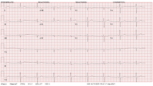
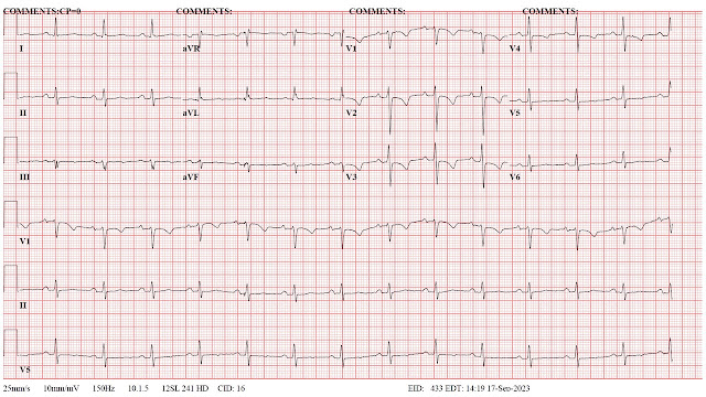
The main implication for me at this point is to cautiously restart my conditioning efforts and see what impact that has on the HRV baseline.  A second question is whether my HRV will approach the pre-ablation baseline.  Electrocardiograms (ECG) may provide some clues in that direction.  I have listed them below for references. Significant changes occurred in the immediate post ablation ECG and the post cardioversion ECG.

An additional thought is whether non linear analysis of the RR intervals would yield more information and easily interpretable graphics. I have used some of these attractor plots in the past and also applied them to single electrode analyses of normal controls and patients with Alzheimer's disease. In terms of ECG analysis - see figure 5 in reference 2. In terms of theory - these attractor diagrams also imply changes in biological complexity at either the structural or functional level - see the diagrams at the bottom of this post


George Dawson, MD, DFAPA


ECG time course (1 -> 5 are in sequence):

1.  Baseline - preop ECG 


2.  Post ablation ECG (following day):

3. Post ablation ECG - note anterior T wave changes thought to be consistent with procedure.


4.  Precardioversion ECG showing atrial flutter at a high rate (day 5 of this arrhythmia; post op day 14).


5. Post cardioversion ECG showing NSR but flipped T waves in V1-V3.


6. ECG follow up 2 weeks after cardioversion showing T wave normalization in anterior leads.


Heart Rate Variability

Some recent recovery in HRV after a long period of low numbers in the 7-37 msec range following ablation and cardioversion.


References:


1:  Fojt O, Holcik J. Applying nonlinear dynamics to ECG signal processing. Two approaches to describing ECG and HRV signals. IEEE Eng Med Biol Mag. 1998 Mar-Apr;17(2):96-101. doi: 10.1109/51.664037. PMID: 9548087.

2:  Nayak SK, Bit A, Dey A, Mohapatra B, Pal K. A Review on the Nonlinear Dynamical System Analysis of Electrocardiogram Signal. J Healthc Eng. 2018 May 2;2018:6920420. doi: 10.1155/2018/6920420. PMID: 29854361; PMCID: PMC5954865.

3:  Aston PJ, Christie MI, Huang YH, Nandi M. Beyond HRV: attractor reconstruction using the entire cardiovascular waveform data for novel feature extraction. Physiol Meas. 2018 Mar 1;39(2):024001. doi: 10.1088/1361-6579/aaa93d. PMID: 29350622; PMCID: PMC5831644.

Monday, September 11, 2023

The Cardiac Ablation


On August 30, 2023, I finally bit the bullet and had a cardiac ablation for atrial fibrillation and atrial flutter. If you are one of those rare readers of this blog you may recall me wrting about it and how it occurred in the first place. I happened to be speedskating 19 years ago on the John Rose Oval and just completed my warm up laps.  I looked at my heart rate monitor and my pulse was 170 BPM.  I pulled up to stretch a little and suddenly my HRM was chirping irregularly and the rate was 240 BPM. I checked my carotid pulse and knew I was in atrial fibrillation. I drove down to the hospital where I was cardioverted with flecainide and metoprolol and have been taking those medications ever since.

In the interim, I have seen a sports cardiologist several times, 5 electrophysiologists (EP), and two general cardiologists as well as my primary care physician and the physicians that cross cover for him. I have also been seen in the emergency department for a heart rate that was down to 25 beats per minute and atrial bigeminy. The physician in the ED thought that I might need a pacemaker, but it turns out that the combination of flecainide and metoprolol can cause significant bradycardia. Once I learned that I started cutting 25 mg tablets into quarters (6.25 mg) and would typically take two of those tablets per day. I also learned that if you take flecainide, you also need to take a beta blocker or a calcium channel blocker to prevent atrial flutter.  Atrial flutter is difficult to diagnose without an ECG because clinically it can seem like sinus tachycardia.  For example, I have had the flu or taken corticosteroids for asthma and developed tachycardia.  When I started running rates of 130 bpm, that seemed a little high for sinus tachycardia.  I decided to get an ECG and it was atrial flutter. I had to figure all of that out, because my initial plan was to taper off metoprolol and that is unrealistic.

At the same time, the combination at times would cause severe bradycardia.  I had a nocturnal heart rate of 35 BPM recorded on a Holter monitor and saw a cardiologist.  We agreed to stay at metoprolol 6.25 mg BID unless there were extraordinary circumstances.  That generally works but my heart rate can still get into the 40s range. That led me to the stage to consider the ablation.  The other factor is that the second EP cardiologist that I saw 15 years ago told me to wait on an ablation because the technology was not good enough. When I saw him this Spring – he thought it had matured and recommended the procedure. He also told me that both the atrial fibrillation and atrial futter could be ablated in a single session rather than two and that was the first time I heard that. 

For about 15 years I have been titrating what most people consider to be microdoses of metoprolol (Physicians typically say: “I have never heard of a dose that small.”) against the flecainide and it has been holding very well. I get about 1 major episode of afib per year that may last 2-3 hours.  I typically take the next dose of flecainide and 12.5 of metoprolol instead of 6.25.   Multiple 24 hr Holter monitors and clinical assessments by cardiologists have not resulted in a better combination.  They were adamant about not increasing the flecainide because of the risk of QRS prolongation and ventricular arrythmias.  There was a consensus to try the ablation – even if the pandemic had persisted.

Researching the procedure followed three lines of evidence.  The first was efficacy and that seems to be a moving target. Conventional wisdom for a long time was that rate control (maintaining a heart rate of < 100 bpm even if you were in atrial fibrillation) and rhythm control (maintaining normal sinus rhythm) produced equivalent results. It turns out that is true only if hemodynamic stability is maintained and for some people it is not.  When that happens, they develop significant symptoms like shortness of breath, lightheadedness, dizziness, chest pain, and can even develop congestive heart failure and renal failure. When all of that is not planned it is riskier to stabilize the person. There is also concern that rate control leads to quality-of-life (QoL) problems associated with both the direct symptoms and indirect symptoms like anxiety about palpitations and the arrhythmia. There seems to be movement in the direction of an attempt to stabilize the rhythm with medication and if that fails try the ablation. There is a QoL rating scale available for atrial fibrillation.  In terms of likelihood of ablating the arrhythmia the frequent quotes are generally 2/3 to ½ of patients, but the data is complicated by the number and intensity of cardiac morbidities.

The second line of evidence was complications and serious complications were noted.  Radiofrequency ablation of arrhythmias in some cases produces a full thickness burn to the heart muscle.  As a result, it can damage adjacent structures including the esophagus and the phrenic nerve.  It can also lead to pericardial effusions and cardiac tamponade. In a very worst-case scenario atrial-esophageal fistula with gas in the left atrium and left ventricle essentially causing an air lock in the pumping mechanism of the heart (4).  

The third line was something I had not considered in the past and that is that atrial fibrillation is progressive. In other words, even if you have good rhythm control with medication, unless something is done to alter the electrical substrate the likelihood of maintaining a normal sinus rhythm after an ablation decreases over time. Accumulating cardiac problems outside of atrial fibrillation can predispose to the condition and make it harder to treat.  

Some additional intangibles were considered. I would like to get back on the ice speedskating. That will take rhythm control and some resilience against exercise induced tachycardia.  Rhythm control is important because atrial fibrillation reduces typical cardiac output by 20-30% based on inadequate filling and pumping cycles due to the irregular heartbeat.   Augmentation of ventricular filling is also adversely affected due to a lack of coordinated atrial contractions.  I am hoping the ablation gets me close to that goal.  There are some theories that interoceptive signaling in the form of accelerated heart rate from any cause can lead to anxiety.  Certainly many people with arrhythmias have anxiety that may seem explainable on a general medical concern basis but there may also be an autonomic component as well as a cognitive component based on the multiple concerns of treating a chronic disorder than can cause stroke and congestive heart failure.    

What has happened so far? I underwent the procedure.  It was 4 hours and 40 minutes in duration from intubation to extubation. The general anesthesia given is shown in the graphics below. The top graphic is the one I made until the official graphical anesthesia record could be located as the second graphic. To do the ablation 4 catheters were placed in the right femoral vein and one in the left. I don’t know the technical details of those catheters only that one is for cryoabalation/isolation of the pulmonary veins in the left atrium, one is for mapping the electrical fields in the surrounding tissue, and one is for a radiofrequency ablation of the a CTI line (cavotricuspid isthmus) in the the right atrium.  That procedure targets atrial flutter.  The plan was do the CTI line ablation first and then puncture the interatrial septum and then enter the left atrium with the cryoablation catheter for the pulmonary vein isolation.  The technical details are more complex since the ablation sites and surrounding areas need to be checked to makes sure that the abnormal conduction sites have been eliminated and no new pathways are evident. The phrenic nerve and esophagus are also checked to make sure there is no damage from ablation that occurs in proximity to these structures. 


Everything seemed to go well during the procedure.  There were no obvious complications just a long time under general anesthesia. Recovery room was uneventful but they decided I needed to stay overnight to monitor bleeding risk from the catheterization site.   That happened when they got me up at the 6-hour mark – blood from the largest site in the right groin dripping onto the floor. More pressure applied and the bleeding stopped and I was discharged the next day.

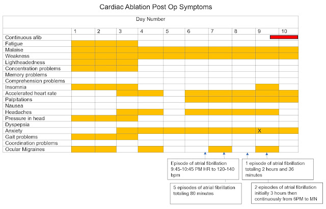
I tried to capture the post-procedure course by in the following graphics.  In clinical practice it was common for me to see people of all ages who had ablations for various arrhythmias. In some cases, they were told to “go home and throw your medications away!” as a result of the ablation.  That may apply to some arrhythmias but not atrial fibrillation. They told me to expect no changes in the medications for 3 months and that I would be taking the same doses of metoprolol and flecainide.  Later at the time of discharge – they told me that in some cases there is a very rocky course until things heal up from the procedure and that it was not uncommon for people to get palpitations and even a return of the rhythm problems.

As noted in the graphics – the course to date has been rocky.  At this point much more atrial fibrillation than I have experienced in the past 16 years and much longer duration.  In my reading about why athletes get atrial fibrillation and the associated experiment work in that area – running sustained high heart rates causes remodeling of the biological substrate of the heart and that makes continued atrial fibrillation more likely. In 16 years, I rarely had an episode that lasted longer than 2 hours and lately more seem to end in less than an hour. As I type this today, I have been in atrial fibrillation for going on 48 hours continuously and just this morning converted to a rapid ventricular response meaning that my ventricular rate is the same as the atrial rate of 150 bpm.  Estimated maximum heart rate for exercise at my age is about 130 bpm.


As can be seen from the graphic there are additional unexpected side effects primary among which is ocular migraines.  An ocular migraine is a typical migraine scotoma without a headache. It starts out as a small shimmering spot or disk in the visual field and slowly expands to a large, jagged, shimmering circle of light. Within about 20 minutes it is gone. Unlike a retinal detachment or stroke there are no deficit symptoms like permanent blind areas in the visual field.  When I asked several staff people about the cause they attributed it to general anesthesia however it is well documented to occur with congenital defects in the atrial septum (patent foramen ovale or PFO) and iatrogenic defects of the septum caused by catheterization into the left atrium (7-10).  Repair of the defect in some cases reverses the headache. About 75% of the iatrogenic atrial septal defects (ASD) spontaneously close by 12 months.  UpToDate put the risk of persistent ASD at 5-20% at 9-12 months (16).

A critical question for anyone contemplating an ablation procedure on a non-acute basis like I did is the post operative course. I was very aware of the low frequency serious and lethal complications, but not the specific about length of time to recovery and what the symptoms might be.  Most people experience significant if not disabling symptoms for months rather than days or weeks following the procedure. That is based on a small study where they did detailed interviews on what happened to the subjects following the ablation (11).  It is available to read online and I would encourage anyone interested in the procedure or knowing more about the procedure to read it.  One of the authors' conclusions is  

“The majority (85%) of the study sample did improve at six months, but the process was much slower and more difficult than expected. Although the symptom burden post-ablation did decrease over the six months, only 50% of subjects (n=10) were symptom-free and off anti-arrhythmic medications at six months.”  (reference 11)  These findings are qualified by the study sample size as well as the possibility of selection bias since the researchers were looking for people who could tolerate the protocol of completing rating scales and lengthy interviews about potential adverse events.  Reference 11 is also very useful in terms for what kind of recovery time to expect - especially in terms of fatigue and more frequent contact with the healthcare system after atrial fibrillation ablation (12).

That is certainly consistent with my experience. Right at this moment I have been in atrial fibrillation or atrial flutter continuously for 48 hours.  My heart rate is 160 bpm at rest.  I am contemplating taking more medication on my own initiative or going to the ED for cardioversion. I am scheduled for a cardioversion in the cardiology clinic on Wednesday September 13 - but I don't know if I can hold off that long.   I guess I am hoping for a break. There are many mitigating factors. Whatever happens tonight – I hope to add more to this post soon.  This is an important topic that has been neglected for too long.

Final qualifier on this post to point out that this is my experience and it does not mean it would be your experience. Much of the sensationalism about medicine in the media is based on oversimplified dichotomous thinking.  Medications, procedures, tests, doctors and even diagnoses are seen as all bad or all good.  Human biology is very complex and there are few if any medical interventions that address that level of complexity. That typically means that over any population there will be an array of outcomes and most of them will not be explainable. That is a hard pill to swallow but that is the state of the art of modern medicine. 


George Dawson, MD, DFAPA


Supplementary 1:  Cardioversion today (9/13/2023) successfully terminated about 90 hours of atrial fibrillation (rates of 70-140 bpm) with atrial flutter (rates of 150-160 bpm).  In terms of the original ablation procedure that is probably more hours of these arrhythmias than I have experienced in the past 19 years.  Normal sinus rhythm has been present for the past 10 hours and vital signs are normal.  What follows is a graphic of the entire process starting with the ablation and ending with the cardioversion. There were multiple episodes of atrial fibrillation before it became continuous with shift to atrial flutter.  During the 90 hours most of the rates were 150-160 bpm.  That is consistent with atrial flutter and may have been associated with a change in medications.


Supplementary 2:

The discussion leading up to the ablation:

 
Image Credit:

Click to enlarge any graphic.

Rottner L, Bellmann B, Lin T, Reissmann B, Tönnis T, Schleberger R, Nies M, Jungen C, Dinshaw L, Klatt N, Dickow J, Münkler P, Meyer C, Metzner A, Rillig A. Catheter Ablation of Atrial Fibrillation: State of the Art and Future Perspectives. Cardiol Ther. 2020 Jun;9(1):45-58. doi: 10.1007/s40119-019-00158-2. Epub 2020 Jan 2. PMID: 31898209; PMCID: PMC7237603.


License : https://creativecommons.org/licenses/by-nc/4.0/This article is distributed under the terms of the Creative Commons Attribution-NonCommercial 4.0 International License (http://creativecommons.org/licenses/by-nc/4.0/), which permits any noncommercial use, distribution, and reproduction in any medium, provided you give appropriate credit to the original author(s) and the source, provide a link to the Creative Commons license, and indicate if changes were made.

Image is used "as is" from the paper and no changes were made.  

Remaining images were all generated by me.


Hat Tip:  Kenneth A. Vatz, MD - a neurology colleague on Twitter who analyzed the scotoma symptoms and directed me to excellent references connecting atrial septal defects to migraines and scotoma especially reference 10. 

Hat Tip:  Medical records staff at Regions Hospital who persevered, located the graphical anesthesia record and mailed it to me on 9/19/2023.  I just incorporated it into this post today.  I have a similar request into the electrophysiology staff so that I can display the actual mapping of this procedure but have been advised that they are less likely to provide these images.     

Update (11/22/2023):  I had a brief (20 min) episode of atrial fibrillation this AM that resolved spontaneously.  It was rate controlled at about 84 BPM.  It is the only arrythmia I have had since the 4 days of atrial flutter early in September.  I notified the clinic and emailed them a tracing of my Kardia ECG.  I have a scheduled appointment next week and it was supposed to be to taper and discontinue the antiarrhythmic medications.  Also seem to correlate with progressively lower HRV numbers despite more vigorous workouts and higher heart rates.


References:

1:  Alobaida M, Alrumayh A. Rate control strategies for atrial fibrillation. Ann Med. 2021 Dec;53(1):682-692. doi: 10.1080/07853890.2021.1930137. PMID: 34032538; PMCID: PMC8158272.

2:  Barbero U, Ho SY. Anatomy of the atria : A road map to the left atrial appendage. Herzschrittmacherther Elektrophysiol. 2017 Dec;28(4):347-354. doi: 10.1007/s00399-017-0535-x. Epub 2017 Nov 3. PMID: 29101544; PMCID: PMC5705746.

3:  Lim HS, Schultz C, Dang J, Alasady M, Lau DH, Brooks AG, Wong CX, Roberts-Thomson KC, Young GD, Worthley MI, Sanders P, Willoughby SR. Time course of inflammation, myocardial injury, and prothrombotic response after radiofrequency catheter ablation for atrial fibrillation. Circ Arrhythm Electrophysiol. 2014 Feb;7(1):83-9. doi: 10.1161/CIRCEP.113.000876. Epub 2014 Jan 20. PMID: 24446024.

4:  Thomson M, El Sakr F. Gas in the Left Atrium and Ventricle. N Engl J Med. 2017 Feb 16;376(7):683. doi: 10.1056/NEJMicm1604787. PMID: 28199804.

5:  Manolis AS. Transseptal Access to the Left Atrium: Tips and Tricks to Keep it Safe Derived from Single Operator Experience and Review of the Literature. Curr Cardiol Rev. 2017;13(4):305-318. doi: 10.2174/1573403X13666170927122036. PMID: 28969539; PMCID: PMC5730964.

6:  Singh SM, Douglas PS, Reddy VY. The incidence and long-term clinical outcome of iatrogenic atrial septal defects secondary to transseptal catheterization with a 12F transseptal sheath. Circ Arrhythm Electrophysiol. 2011 Apr;4(2):166-71. doi: 10.1161/CIRCEP.110.959015. Epub 2011 Jan 19. PMID: 21248245.

7:  Kato Y, Hayashi T, Kato R, Takao M. Migraine-like Headache after Transseptal Puncture for Catheter Ablation: A Case Report and Review of the Literature. Intern Med. 2019 Aug 15;58(16):2393-2395. doi: 10.2169/internalmedicine.2519-18. Epub 2019 Apr 17. PMID: 30996181; PMCID: PMC6746642.

8:  Hoshina Y, Iijima H, Kubota M, Murakami T, Nagai A. Case of atrial septal defect closure relieving refractory migraine. Clin Case Rep. 2022 Nov 6;10(11):e6484. doi: 10.1002/ccr3.6484. PMID: 36381060; PMCID: PMC9637252.

9:  Azarbal B, Tobis J, Suh W, Chan V, Dao C, Gaster R. Association of interatrial shunts and migraine headaches: impact of transcatheter closure. J Am Coll Cardiol. 2005 Feb 15;45(4):489-92. doi: 10.1016/j.jacc.2004.09.075. PMID: 15708691.

10:  Schwedt TJ. The migraine association with cardiac anomalies, cardiovascular disease, and stroke. Neurol Clin. 2009 May;27(2):513-23. doi: 10.1016/j.ncl.2008.11.006. PMID: 19289229; PMCID: PMC2696390.

11:  Wood KA, Barnes AH, Paul S, Hines KA, Jackson KP. Symptom challenges after atrial fibrillation ablation. Heart Lung. 2017 Nov-Dec;46(6):425-431. doi: 10.1016/j.hrtlng.2017.08.007. Epub 2017 Sep 18. PMID: 28923248; PMCID: PMC5811184.

12:  Wood KA, Barnes AH, Jennings BM. Trajectories of Recovery after Atrial Fibrillation Ablation. West J Nurs Res. 2022 Jul;44(7):653-661. doi: 10.1177/01939459211012087. Epub 2021 Apr 26. PMID: 33899608; PMCID: PMC8801207.

13:  Björkenheim A, Brandes A, Magnuson A, Chemnitz A, Svedberg L, Edvardsson N, Poçi D. Assessment of atrial fibrillation–specific symptoms before and 2 years after atrial fibrillation ablation: do patients and physicians differ in their perception of symptom relief?. JACC: Clinical Electrophysiology. 2017 Oct;3(10):1168-76.

14:  Dorian P, Angaran P. Symptoms and Quality of Life After Atrial Fibrillation Ablation: Two Different Concepts. JACC Clin Electrophysiol. 2017 Oct;3(10):1177-1179. doi: 10.1016/j.jacep.2017.06.007. Epub 2017 Sep 13. PMID: 29759502.

15:  Steinbeck G, Sinner MF, Lutz M, Müller-Nurasyid M, Kääb S, Reinecke H. Incidence of complications related to catheter ablation of atrial fibrillation and atrial flutter: a nationwide in-hospital analysis of administrative data for Germany in 2014. Eur Heart J. 2018 Dec 1;39(45):4020-4029. doi: 10.1093/eurheartj/ehy452. PMID: 30085086; PMCID: PMC6269631.

16:  Levy S.  Overview of catheter ablation of cardiac arrhythmias.  In: UpToDate, Connor RF (Ed), Wolters Kluwer. (Accessed on January 17, 2023)