Showing posts with label ECG. Show all posts

Friday, September 22, 2023

Heart Rate Variability


I have been following heart rate variability (HRV) on my watch and three different apps for the past several years. HRV is defined as the slight variations between R waves in the standard ECG recording.  I have included an example below, illustrating the R-R’ intervals (or RRI) and how they might vary over time.

Since HRV became widely available as a measurement off a watch that is commonly worn by millions of people, the research on this measurement and the variable studied has increased significantly.  For my purposes – HRV is thought to be an indicator of heart health and conditioning and possibly a marker of overtraining – but advice about that varies significantly. Some studies have shown that decreased HRV is associated with an increased risk of arrhythmias.  My recent cardiac ablation and cardioversion seemed to present an ideal situation for further study. 

Before getting into those details the physiology of HRV needs to be considered. The dominant heart rhythm of a normal heart is determined by the sinoatrial (SA) node. This node contains a population of spontaneously depolarizing cells that determine the rhythm and rate of the heartbeat. In addition to the neurophysiology of that cell population several additional factors affect both the rate and HRV.  Primary among them is autonomic innervation from both the sympathetic and parasympathetic systems and their effect at the SA node. Parasympathetic fibers from the vagus nerve modulate slower firing through the neurotransmitter acetylcholine (ACh). Sympathetic fibers increase the rate of firing through the neurotransmitter norepinephrine (NE).  NE has a longer half-life than ACh, but vagal tone is thought to be the most significant determinant of HRV.  That is in line with several clinical observations including lower baseline heart rates in conditioned athletes and higher heart rates in people with less conditioning or in stressful situations.

What happened to my heart rate and HRV during the recent cardiac ablation for atrial fibrillation and subsequent cardioversion?  To answer that question, I had to figure out how to get the data off my Apple Watch 5.0.  The only approach I could find was to downloaded all of the collected Health App data as a CSV file and then plot it in Excel.  There are some online sites that you can download the data to and then use the remote software for plotting, but I preferred to retain control over the data. If you decide to do that and have several years of data like I did – it takes a long time.  It took about 5 hours in my case to download about 1G of data to a zip file.  From there it is easy to open that file with Excel or other software and do the plots. A useful addition to the Health App would be able to download specific time intervals.

I have done 2 plots so far based on average daily HRV and hourly HRV as shown below.


 The plots are interesting because it clearly shows an effect from the ablation, a 96-hour period of atrial fibrillation and atrial flutter, and the cardioversion. At the minimum the baseline HRV drops to a different baseline after the ablation. That is followed by a significant spike with the recurrence of afib/flutter.  And then there is a return to the lower baseline after the electrical cardioversion.  I rarely had any significant episodes over the course of a year and whenever I went back and reviewed HRV it was not significantly changed. Since all those episodes were typically less than 2 or 3 hours it may not have been long enough to see an HRV effect.  Conversely spikes of 50-100 msec in the HRV recording were common and not associated with arrhythmias. In the case of the post ablation period the sustained rates were associated with spikes, but since atrial flutter is regular, the associated R-R’ intervals probably showed a more characteristic HRV.

I would expect to see an increase in vagal tone and therefore HRV just related to the sustained high rates over 4 days. If increased vagal tone correlates with increased HRV that does not seem to be the case in these graphs. The graphs also seem to indicate to me that there may be a structural element to HRV – either in the anatomical configuration of the conducting cells, their altered physiology, or a combination.

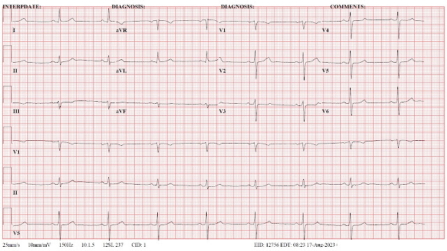
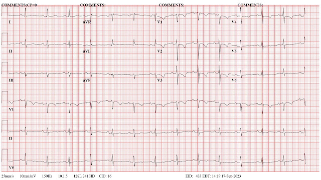
The main implication for me at this point is to cautiously restart my conditioning efforts and see what impact that has on the HRV baseline.  A second question is whether my HRV will approach the pre-ablation baseline.  Electrocardiograms (ECG) may provide some clues in that direction.  I have listed them below for references. Significant changes occurred in the immediate post ablation ECG and the post cardioversion ECG.

An additional thought is whether non linear analysis of the RR intervals would yield more information and easily interpretable graphics. I have used some of these attractor plots in the past and also applied them to single electrode analyses of normal controls and patients with Alzheimer's disease. In terms of ECG analysis - see figure 5 in reference 2. In terms of theory - these attractor diagrams also imply changes in biological complexity at either the structural or functional level - see the diagrams at the bottom of this post


George Dawson, MD, DFAPA


ECG time course (1 -> 5 are in sequence):

1.  Baseline - preop ECG 


2.  Post ablation ECG (following day):

3. Post ablation ECG - note anterior T wave changes thought to be consistent with procedure.


4.  Precardioversion ECG showing atrial flutter at a high rate (day 5 of this arrhythmia; post op day 14).


5. Post cardioversion ECG showing NSR but flipped T waves in V1-V3.


6. ECG follow up 2 weeks after cardioversion showing T wave normalization in anterior leads.


Heart Rate Variability

Some recent recovery in HRV after a long period of low numbers in the 7-37 msec range following ablation and cardioversion.


References:


1:  Fojt O, Holcik J. Applying nonlinear dynamics to ECG signal processing. Two approaches to describing ECG and HRV signals. IEEE Eng Med Biol Mag. 1998 Mar-Apr;17(2):96-101. doi: 10.1109/51.664037. PMID: 9548087.

2:  Nayak SK, Bit A, Dey A, Mohapatra B, Pal K. A Review on the Nonlinear Dynamical System Analysis of Electrocardiogram Signal. J Healthc Eng. 2018 May 2;2018:6920420. doi: 10.1155/2018/6920420. PMID: 29854361; PMCID: PMC5954865.

3:  Aston PJ, Christie MI, Huang YH, Nandi M. Beyond HRV: attractor reconstruction using the entire cardiovascular waveform data for novel feature extraction. Physiol Meas. 2018 Mar 1;39(2):024001. doi: 10.1088/1361-6579/aaa93d. PMID: 29350622; PMCID: PMC5831644.

Saturday, September 21, 2019

Cardiac Screening In Psychiatric Patients


There is a paper that just came out (1) that I consider a must-read for all psychiatrists.  Some experts might qualify that and say that it is only necessary to know the subjects if you are treating the medically ill, the elderly, pediatric patients, patients with cardiovascular disease, or patients with cardiac risk factors. The problem with those qualifications is that you have to know everything in this paper (and others) in order to make that determination. Beyond that you have to been trained in how to determine if your patient is seriously ill or not. In the case of all medical specialists, serious illness generally means treatment by another specialist or in a more intensive setting. For that reason, the cardiac aspects of care in this paper are required knowledge.

Three of the authors of this paper are cardiologists, two are psychiatrists, and one is a clinical pharmacist. They have produced a very practical document on identifying problems with tachycardia, QTc prolongation, sudden cardiac death, myocarditis, and dilated cardiomyopathy.  They provide very specific endpoints and suggest some basic intervention that can be done before the patient is referred to cardiology. Examples would be assessment of tachycardia, suggested treatment thresholds, common treatment interventions like beta-blockers and calcium channel blockers, and referral to cardiology if there is a progression to other cardiac symptoms, nonresponse to the initial therapy, or an arrhythmia beyond sinus tachycardia. They provide similar guidance on the other common conditions and relate them to second-generation antipsychotics (SGAs). 

All of the authors are from the United Kingdom.  I am not familiar with the standard settings for practicing psychiatry in the UK, but in the US there is a high degree of variability. For example, in practicing on inpatient settings it is not a problem to order ECGs or even stat ECGs. Echocardiograms and other imaging studies of the heart are easily obtained as well as cardiology consultation. In a previous inpatient setting where I practiced, I requested a cardiology consultation for a young woman with a QTc of 520 ms who required treatment with antipsychotic medications. She was seen immediately and an electrophysiology study was done. After that study I was advised by cardiology that I could safely treat the patient with olanzapine. At the other end of the spectrum, I know there are psychiatrists reading this who have no access ECGs, medical testing, or cardiology consultants. They are often practicing in an office that lacks a sphygmomanometer or staff routinely checking patient vital signs. Many of those office settings are essentially nonmedical and any psychiatrist practicing there - would need to bring in their own equipment and probably take their own vital signs.  A basic standards would be that every practice setting for psychiatry in the country should have the tools to make the measurements recommended in this paper, but I am not aware that any standard like that exists.

The second obstacle to realizing these guidelines is the way electronic health records (EHR) are set up. Major organizations and the EHR companies themselves produce templates that are typically designed for business purposes rather than medical quality. A visit to a psychiatrist in that organization results in that template being filled out with a business rather than a clinical focus. In other words, sections of bullet points are completed based on what coders believe will capture the necessary billing from insurance companies. One of the key sections is often the review of systems (ROS). Because these documents are not designed by physicians and there are no uniform standards, a functional review systems is often not there. In the case of cardiac symptoms, there needs to be a clear section that encompasses all the symptoms described by the authors in this paper. As an example, take a look at the cardiac symptomatology that I recorded in this post and the modified extended review of systems that I typically ask patients about.

Any inpatient or outpatient assessment done by a psychiatrist should include a thorough medical history, a review of systems that is focused on medical rather than psychiatric systems, a set of vital signs including noting whether or not the cardiac rhythm or pulse is regular or irregular and a further description of the irregularity.  A focal exam for additional heart and lung findings and determination of pulses and peripheral edema may be indicated.  The take home point is that this history taking combined with a few additional findings should be all that is necessary to order further tests like an ECG, refer for an acute assessment, or refer to a primary care physician or cardiologist for further assessment.  If the patient is being followed for the metabolic complications of SGAs, there may already be a fasting blood glucose, and lipid profile in the chart to assess additional risk factors.  Over the years I have also found that recording a theory about why I think the patient is symptomatic is also very useful.  In my practice that has ranged from medication side effects to an acute myocardial infarction.

With those issues we can proceed to consider the assessments and treatments recommended by this group. I am not going to repeat the content of the paper here.  I recommend that any interested psychiatrist or psychiatric resident get a copy of this paper and study it in detail if you are not already familiar with the concepts. I will list a few of the points that I found to be interesting after doing these assessments for a long time.

Tacycardia is a common problem in psychiatric patients and the population in general. Here the authors were focused on tachycardia as a side effect of SGAs and haloperidol.  They produced a table showing the incidence of tachycardia across a number of SGAs and haloperidol and illustrate that clozapine by far has the highest prevalence of tachycardia. In the table haloperidol, asenapine, and sertindole at the lowest incidence of tachycardia at about 1%. They point out this problem is generally self-limited but it suggests a number of investigations that should be considered before monitoring for improvement over time. The recommended treatments (bisoprolol, ivabradine) are not recognizable medications for physicians in the US. In the US, beta-blockers are commonly used. They suggested treatment is predicated on whether patients are symptomatic or not with palpitations. Although UpToDate describes sinus tachycardia as a benign condition with no worse outcome than a control group, this tachycardia is drug-induced. My main concern with persistent drug-induced tachycardia is tachycardia induced cardiomyopathy. My other concern is that common causes of tachycardia in the patients I see include excessive use of caffeine (alcohol, or other substances), deconditioning, and sleep deprivation. Establishing a baseline prior to any of these prescriptions is important.  There is always a lot of debate about whether or not electrocardiogram should be done. I agree with the authors that the ECG is an inexpensive screen and should be done to make sure that it is a sinus rhythm. Another bit of information that may not be available is whether the pulse is irregular or not. Many clinics have automatic blood pressure and heart rate measuring devices and not all of them make that determination.

The section on the QTc interval was interesting because the authors provide very clear guidance on measuring QTc, the problems with that measurement, and very clear guidelines on what to do about that measurement. They cite the threshold for stopping or reducing treatment with QTc prolonging agents as an interval greater than 500 ms or relative increase of greater than 60 ms. They also use the American Heart Association definitions of prolonged as QTc > 450 ms in men and > 460 ms in women.  They point out that the most common calculation of QTc (Bazett’s formula) overcorrects heart rates greater than 100 BPM and they suggest that other formulae may be used for that situation. Like many psychiatrists I have ordered hundreds of ECGs for determining baseline cardiac conduction. The vast majority have been normal. The ones that were not - were typically unrelated to the medication I was prescribing. Many conduction abnormalities were related to increasing age and latent cardiac problems. The other common scenario where I am concerned about cardiac conduction is polypharmacy. It is possible for a person to be taking multiple medications for psychiatric indications - all of which may affect cardiac conduction. The drug interaction software for most EHRs as a very low threshold for this type of interaction.

The myocarditis section of this paper was very interesting. In Table 1 - the authors included prevalence figures for myocarditis in the same table where they documented the prevalence of tachycardia for each medication. The figures are based on isolated case reports. The review the controversy about clozapine and widely variable reports of incidence. The incidence quoted for Canada and the USA was 0.03%. Different criteria used to diagnose myocarditis was considered an important point of variance. A set of clinical criteria is provided in the paper as well as when to refer to a cardiologist. In addition to the ECG, serum troponin, C-reactive protein, echocardiogram, and cardiac MRI are considered. The referral indicators included elevated troponin, CRP, and abnormalities at echocardiogram. My interpretation is that psychiatrists in the US who have access to those measures and ready access to cardiologists could potentially use those markers. The most reasonable approache is to be able to recognize the symptoms of myocarditis clinically and be able to refer the patient to cardiology were most of the testing could occur. The clinical description of myocarditis in the paper sounded very similar to typical viral myocarditis with chest pain, dyspnea, flu-like illness, fever, and fatigue. These are nonspecific symptoms especially during influenza season. The clinician has to have a high index of suspicion based on treatment with clozapine. The paper contains an ECG tracing of saddle -shaped ST elevation considered to be a finding consistent with myocarditis. It was visible in most leads.

The approach to dilated cardiomyopathy was very similar in terms of recognizing the symptoms of congestive heart failure and the necessary investigations. There was guidance and when to request an echocardiogram based on BNP and in NT-ProBNP measures as well as when referral to cardiology was indicated. The standard of care in the US is the psychiatrist recognizing what is happening but not treating dilated cardiomyopathy. In most clinical with limited resources, this is a good reason to have a referral relationship with a primary care clinic - especially one that can do the testing on site. There are many primary care and even urgent care clinics that cannot do the testing suggested in this paper.

In the case of myocarditis and dilated cardiomyopathy, the question of whether a patient should be re-challenged if they need the offending medication and their underlying cardiac condition has improved. The authors suggest close consultation with a cardiologist at that point. Given the data my own practice has been to not re-challenge with the offending medication but to try a different treatment modality. The concern in the article is that the patient’s ability to function from a psychiatric standpoint may require use of that specific medication. I do not think that enough is known about the outcome of either condition to resume the original medication, but if favorable outcome studies or case reports exist, I might revise that opinion.

All things considered this is an outstanding article on the cardiotoxicity of SGAs. The graphics in the paper also excellent with management flow diagrams and well-designed tables.  The authors restate that cardiotoxicity is very low.  It is the job of every psychiatrist who prescribes these medications and others to make sure that patients are monitored for these complications. There is always a question of what constitutes adequate informed consent when we are talking about a potential complication rate of 0.03%. At that level it is certainly possible that many psychiatrists have never seen these complications and never will. I think it is reasonable to let people know that medications they are taking can cause rare but potentially serious side effects including death. The informed consent issue was not touched on in the paper but a day-to-day practice it is an important one.  From a practical standpoint I generally advise people that if they are taking a medication with rare but potentially life threatening side effects, they have to take all physical symptoms seriously. Physical symptoms cannot be attributed to common explanations like colds, the flu, or gastroenteritis.

This paper had a very specific focus and it did not touch on the other metabolic and neurological complications of these medications that require additional screening.  One of the reasons I posted my ROS document on this blog was to make ti easy for any clinic or psychiatrist to build their own template with the relevant questions needed for their own patient population. 

For some psychiatrists and clinics the work in cardiac screening just got a lot harder.  For others who have been doing all of this for decades - there will be very little difference.


George Dawson, MD, DFAPA


References:

1:  Sweeney M, Whisky E, Patel RK, Tracy DK, Shergill SS, Plymen CM.  understanding and managing cardiac side effects of second-generation antipsychotics in the treatment of schizophrenia. Br J Psych Advances 2019: 1-15.

2:   Patel RK, Moore AM, Piper S, Sweeney M, Whiskey E, Cole G, Shergill SS, Plymen CM. Clozapine and cardiotoxicity - A guide for psychiatrists written by cardiologists. Psychiatry Res. 2019 Jul 24:112491. doi:10.1016/j.psychres.2019.112491. [Epub ahead of print] Review. PubMed PMID: 31351758.


Tuesday, March 31, 2015

No Information From The EHR - An Ongoing Problem


Like most physicians - I like the concept of an electronic health record (EHR).  It is just that the real EHR as it exists is a far cry from the concept.  The proponents of the current EHR,  especially those who want it mandated by legislative activity continue to brag about the savings and all of the benefits.  Any physician looking for information or an ability to enter and move information without ending up in a click fest of mouse clicks knows the reality.  Any physician looking for a note that reflects an intelligent conversation between a physician and a patient is also left wanting.  Reading the electronic or printed out version of the EHR usually results in very choppy documentation.  Lists that are the result of not very intelligent coding by EHR IT engineers, notes produced strictly to meet billing and coding bullet points, and notes produced because they could be rapidly compiled with features like smart text.

All of this can be a nightmare for a compulsive physician like myself who wants to use all of the relevant information in patient care.  My career has been treating patients with complex medical conditions who are also on complicated combinations of medications.  Many have known heart disease and take combination of medication that can adversely affect their cardiovascular status and interact with psychiatric medications that I prescribe.  All of that needs to be considered.  Since ziprasidone (Geodon) hit the market in 2001, psychiatrists have been preoccupied with the QTc interval.  The QTc interval is the electrical interval that corresponds to the contraction and relaxation of the left ventricle.  In cases where this interval is too long it predisposes the patient to ventricular arrhythmias some of which are potentially fatal.   The FDA had a warning on ziprasidone about the potential for QTc prolongation and subsequently came out with warnings about citalopram.  In the course of clinical practice, many psychiatrists had already encountered this issue with older antipsychotic medications and tricyclic antidepressants.  The FDA makes these pronouncements but gives physicians no guidance on what to do about the clinical situations.  I have a practice of looking at ECGs and any Cardiology evaluations that have been done.  That is the only way the QTc interval can be determined and even then there are various factors that can affect it.

Rather than order an ECG, I will ask whether they have already been done and get the patients consent to have them faxed to me.  That result is frequently disappointing, especially in the case of the EHR.  I will often get a series of cryptic sheets, that look like a sparsely populated medical record.  There are often no coherent notes from physicians or if they are there, they do not contain standard information that I am looking for.  I have never seen an ECG tracing contained in these stack of records.  The best I can hope for is a brief note that lists an impression like "NSR - no acute changes."  An added bonus would be an actual description of the critical intervals.  For the tracing at the top of this page it would say:  "PR interval - 164 ms; QRS duration - 100 ms; QT/QTc - 434/415 ms."  That is really all of the information I need to know.  But the most important issue with the EHR is that all of this visual information is usually lost, unless I submit a second or third request and it usually has to say "send me the ECG tracing."  The medium that purports to provide a lot of information to physicians and put it at their fingertips is a bottleneck.  By the time I see the information I need to see, it is not necessary.  I have moved on and not recommended a treatment that I could have recommended if the ECG was normal.  That practice has been reinforced by getting an ECG after the fact and realizing that not only was there a prolonged QTc interval, and it was read that way by a Cardiologist but reported as "normal" in the EHR.

I will be the first to admit that there is minimal evidence that my tight QTc surveillance has saved any lives.  But my threshold is really to prevent any complications.  I am not treating acute heart conditions.  I am trying to make sure that I don't cause any by the medications that I prescribe, by ignoring a critical drug interaction, or by not recognizing the significance of a patients physical illness and how it needs to direct the therapy that I prescribe.

That doesn't end at ECGs.  I would throw in imaging studies (CT and MRI), EEGs, and even routine labs.  If the EHR is supposed to convey the maximum information why wouldn't all of the visual information of an episode of care be included?  Why can't all of the brain imaging studies be sent along as a disk or e-mailed to me?  Why do I have to read a 200 page fax and try to reconstruct all of the lab results  in a coherent manner that are spread randomly across those pages so that I know what happened in the hospital?

The EHR as it currently exists is a tremendous burden to physicians.  It takes far too long to enter data and quality notes about care are rare.  If you happen to lack online access to the program where the record is constructed, good like trying to piece together the information that you need for clinical decision-making.  Politicians are good with ideas, but none of them seems to be aware of the real problems that exist in these systems.  Despite that lack of knowledge they continue to insist on the wide implementation of these systems and that is really a tax on physicians that is being used to subsidize the development of EHRs and fund this industry.

Hopefully that will pay off someday, but the current problems have been there for at least a decade and there are no signs that they will be going away soon..


George Dawson, MD, DFAPA  

Friday, November 23, 2012

Mayo Clinic Counterpoint to FDA on Citalopram

The Mayo Clinic came out with their recommendations on what to do about the FDA's warning about citalopram.  By their own description they are more liberal with regard to their citalopram recommendations and more conservative regarding escitalopram.  I have previously reviewed the problem here and concluded that there is really a lack of data available on the likelihood of electrocardiogram abnormalities during normal clinical use and if citalopram is as cardiotoxic as the FDA is describing it - we should treat it more like a standard antiarrhythmic drug and used flecanide as an example.

For all practical purposes that would include baseline ECGs, ECGs at the max dose and taking it up one more level from either the Mayo Clinic or the FDA - a stress test looking for QTc prolongation at higher heart rates.  The other elements in the Mayo recommendations based on history and physical examination and expecting some physician knowledge of drug metabolism are fairly standard.  I thought it was interesting that they did not mention checking plasma levels of the drug especially in complex cases (eg. a patient with cirrhosis) who only responds to higher than recommended doses of the drug.  Regarding the statements:  "Selective serotonin reuptake inhibitors cannot simply be substituted for one another, not even escitalopram for citalopram."  That is generally true and where are these guys in the battle against PBMs saying that these drugs are all equivalent?  I have not found any patient that responded selectively to citalopram and not escitalopram.  I have generally been able to convert patients to an equivalent amount of escitalopram the next day.

Both the Mayo Clinic and the FDA are silent on molecular approaches to solving this problem and screening patient for potential risk before they are started on either drug.  The Mayo Clinic offers testing for cytochrome P450 genotypes.  The genetic basis for hereditary prolonged QTc intervals has been a hot topic of research over the past decade.  It is probably time to expand the search for additional genotypes that place people at risk during specific drug therapies.  Until then we have only very approximate methods of determining the at - risk population and keeping them safe and the Mayo recommendations are more reality based than the FDA.

I think it would also be possible to estimate the risk associated with taking citalopram across the entire population.  In fact, at this point the FDA seems to have the data to estimate the risk of any QTc effect at all to the risk of torsade de pointes - the most significant arrhythmia.  I think it is very important for patients making the decision to have this number and if I can provide numbers on rare but serious antidepressant complications like serotonin syndrome, a federal agency with more perfect information and no patient care responsibility can do better.

George Dawson, MD, DFAPA

Sheeler RD, Ackerman MJ, Richelson E, Nelson TK, Staab JP, Tangalos EG, Dieser LM, Cunningham JL. Considerations on safety concerns about citalopram prescribing. Mayo Clin Proc. 2012 Nov;87(11):1042-5.

FDA Drug Safety Communication: Revised recommendations for Celexa (citalopram hydrobromide) related to a potential risk of abnormal heart rhythms with high doses.


Sunday, April 29, 2012

Does the FDA discriminate against antidepressants?


The FDA came out with a new warning on citalopram on 3/28/2012.  The main point of the warning is that citalopram may lead to electrocardiogram changes that can be associated with an abnormal heart rhythm or arrhythmia that is potentially fatal.  The specific change is prolongation of the QTc interval or the interval that correlates with the total duration of ventricular activation and recovery.

Citalopram is a widely used antidepressant medication and it widely used for three reasons.  It is not likely to have a lot of interactions with other drugs.  Citalopram figured prominently in the STAR*D algorithm from the largest study done on enhancing antidepressant effectiveness.  A third reason is that it is a generic medication and it is very inexpensive.  Psychiatrists have broad experience with the drug and the general experience is that it is well tolerated with little toxicity.

Flecainide is a Type IC antiarrhythmic agent indicated for the prevention of paroxysmal atrial fibrillation (AF), paroxysmal supraventricular tachycardia (PSVT), and the prevention of life-threatening ventricular  arrhythmias like sustained ventricular tachycardia. The FDA warnings on the drug include proarrhytmic effects and excess mortality.  The excess mortality was directly observed in a clinical trial done to suppress ventricular arrhythmias.

The black box warnings for each drug listed below are directly from Medline:


Looking at the safety concerns for both medications - important differences emerge.  First, the FDA recommends maximum doses for the citalopram not just for the a maximum dose for adults but in specific conditions including aging.  Searching the FDA web site shows exactly 25 references for safety concerns of flecainide and none of them contain that level of information.  Second, the citalopram warning shows a table of QTc interval changes by dose for both citalopram and escitalopram.  There is no information in FDA documents (that I could find) for flecainide even though it is widely accepted that flecainide causes dose related changes in not just the QTc interval but also the QRS and PR intervals  along with a host of additional effects on cardiac pacemakers and conduction.  The  overall tone of the release is  that citalopram is a potentially cardiotoxic drug.  Third, the ECG monitoring recommendations are not internally consistent.  The absolute cut off of a QTc interval of 500 ms is highly unlikely - even in cases where the patient is taking 60 mg per day or more of citalopram.  It is also unlikely that the QTc intervals in the citalopram warning will lead to a QTc interval of greater than 500 ms.  This will result in tens of thousands of ECGs done because that is the only way that the QTc interval can be determined.

The black box warnings and the recently issued warning all considered, serious questions are raised relative to drugs with known cardiotoxicity and the whole issue of QTc warnings in all psychiatric drugs.  Certainly nobody wants a rare severe complication as a result of a prescription medication but can it really be avoided?  What good would ECG screening do?  There have not been any trials to address that issue of whether all patients taking citalopram need baseline ECGs.  All the patients taking flecainide have probably had multiple ECGs done that indicate a possible need for treatment but there is little guidance on the ECG issue.  In many patients taking flecainide, patients get serial ECGs and they do exercise stress tests to rule out proarrhythmic effects.  Are the same precautions needed for patients on citalopram?

Are the thresholds for treatment different given the fact that flecainide caused increased mortality during clinical trials and citalopram did not?  There would be an argument that flecainide is used to treat life-threatening arrhythmias, but the other indication is for prevention of atrial fibrillation and atrial fibrillation is not a life threatening arrhythmia.  With regard to the seriousness of the diagnosis, major depression carries a lifetime mortality of 10%.  Finally, where is the table on the relationship between flecainide dose and QTc prolongation like we see for both citalopram and escitalopram?  Is it possible that flecainide has more of an effect throughout the dosage range than citalopram?

These are serious questions given that I have already established that there is a significant bias in the media against psychiatry, psychiatrists and psychiatric medications.  The most recent FDA warning has created a lot of anxiety for psychiatrists and any patient taking citalopram.  The majority of those patients are being seen by primary care physicians.
  
If citalopram is that cardiotoxic, let's see the evidence and let's see how it compares to a medication with known cardiotoxicity.  Let's have the same level of warning for both medications and some concrete ideas about what needs to be done to manage that risk.

George Dawson, MD, DFAPA